

Ghyass Jutt: Can You Identify a Euploid Blastocyst Under the Inverted Microscope?
Ghyass Jutt, Embryologist at Sakina International Hospital at the Andrology Institute of Pakistan (STAR ICSI), shared on LinkedIn:
“Can you identify a Euploid Blastocyst under the inverted microscope?
The short answer: Not definitively.
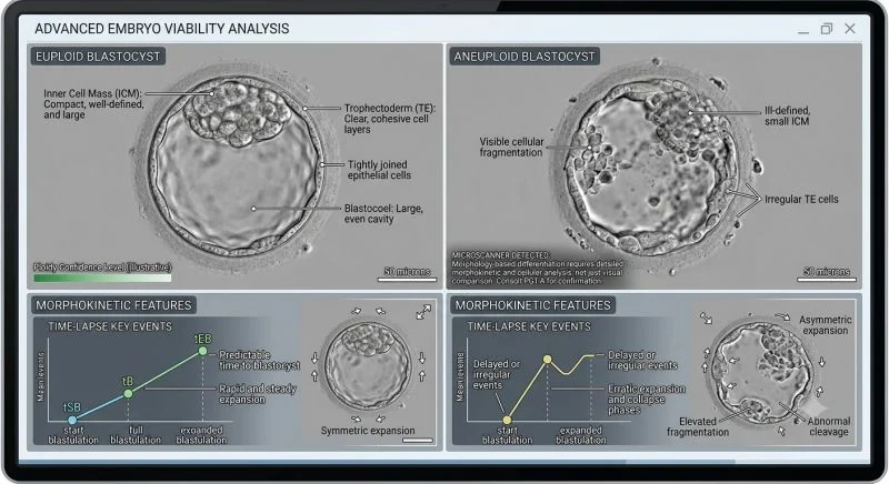
While high-grade morphology and tight cellular cohesion statistically correlate with normal chromosomal status, visual assessment is a proxy, not a diagnostic tool. A visually perfect 5AA blastocyst can be aneuploid, whereas a lower-grade CC embryo can result in a healthy euploid live birth.
Morphological and Morphokinetic Trends
When grading and prioritizing Day 5 or Day 6 blastocysts, embryologists evaluate several key statistical markers:
Euploid Indicators:
- Inner Cell Mass (ICM): Compact, prominent, and densely packed with uniform cells.
- Trophectoderm (TE): Cohesive epithelium with clear, tightly joined cells forming a continuous wall.
- Dynamics: Steady, symmetric blastocoel cavity expansion with on-time developmental milestones.
Aneuploid Red Flags:
- Structural Degeneration: Small or fragmented ICM alongside sparse, uneven TE cells.
- Developmental Delays: Prolonged time to blastulation (delayed into Day 6 or Day 7).
- Morphokinetic Perturbations: Erratic cleavage events, elevated fragmentation, or repeated collapse and re-expansion cycles.
Why Optical Microscopy Reaches Its Limit:
- Mosaicism: Visual inspection cannot resolve cell-level chromosomal variations across trophectoderm and ICM lineages.
- Normal Mitotic Function in Aneuploidy: Aneuploid cells frequently undergo normal mitosis and fluid pumping, creating visually top-grade blastocysts.
- Embryonic Self-Correction: Blastocysts can sequester or extrude aneuploid cells into the perivitelline space while leaving a normal inner cell mass.
High-power light microscopy remains indispensable for embryo selection and prioritization, but trophectoderm biopsy with PGT-A (NGS) remains the sole diagnostic standard for verifying chromosomal status.
How heavily does your laboratory weigh Day 5 vs. Day 6 expansion kinetics when prioritizing transfers in non-PGT-A cycles? Let’s share insights below!”
Stay informed with the latest updates in fertility and reproductive medicine on Fertility News.
-
Nov 22, 2026, 17:01Insights from ISFP 2026
-
Nov 22, 2026, 16:17Khalil Faaed: The Dark Center of the Oocyte
-
Nov 22, 2026, 16:10Jyoti Gupta: Proud to Join Cloudnine Fertility, Golf Course Road, Gurugram, as Director
-
Nov 22, 2026, 15:45Francine Freitas Fernandes: 3D and 4D Hysterosalpingo-Contrast Sonography
-
Nov 22, 2026, 15:25New RBMO Study Links Microfluidic Sperm Sorting to Higher Blastocyst Formation and Lower Miscarriage Rates
-
Nov 22, 2026, 15:21Michelle Mason: Pleased to Attend the Annual SWIGS Conference with the Care Fertility Cardiff Team
-
Nov 22, 2026, 15:18Rajeev Agarwal: The Lining Is 6.5 mm – Will the Embryo Transfer Fail?
-
Nov 22, 2026, 15:11The Automated IVF Laboratory Lecture by Jacques Cohen at the 5th Narture – Conceivable Life Sciences
-
Nov 22, 2026, 15:07Laura Rienzi: Same Message, 2 Slides – Which One Would You Rather See at a Congress?