

Revolutionizing Uterine Drug Development with Dynamic Imaging – Fidēs Imaging
Fidēs Imaging shared a post on LinkedIn:
“Is it time to bring dynamic functional imaging into mainstream uterine drug development?
Drug trials in uterine disorders consistently show higher failure rates than many other therapeutic areas.
Why?
Underinvestment in R and D plays a role. So does disease complexity. But one critical issue stands out: unclear and incomplete functional endpoints.
The physiology of the female reproductive system is complex, and a clear understanding of the endometrial cycle and the motion of the uterus in healthy/diseased states must be central to any research in this space. This is not an entirely novel concept in academic research, but now needs to permeate drug development.
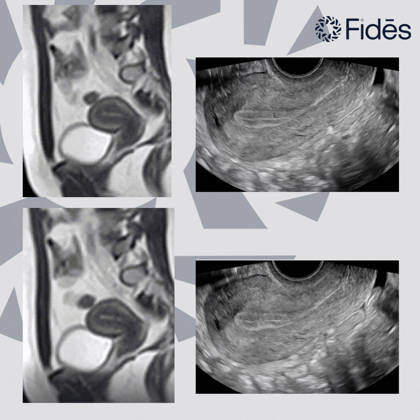
Standard imaging techniques still focus on static images and scores. These snapshots focus on structure, not function. Given that most drug development is concerned with function, it is clear that we’re missing a key dimension – movement.
Across the menstrual cycle, the uterus is dynamic, from menstruation (where we see fundocervical waves) through the follicular phase (involving cervico-fundal peristalsis) to the luteal phase (reduced peristalsis and changes in the direction of contractions) and finally ovulation (increased frequency and strength of contractions), to start the process again.
Understanding this complex pattern of uterine movements, and how they differ under different conditions: endometriosis, adenomyosis, infertility, pain, etc – needs a better understanding of function/movement of the organ. The academic field of research is somewhat thin here, but provides some interesting conclusions ‘(to) support the hypothesis that uterine contractility … plays a central role in fertility and that gynaecological pathologies affect the quality and quantity of these contractions’.
Extending that thought: if motion changes, what does that mean for pain, fertility, implantation, or long-term function?
What Fidēs brings to clinical trials is unparalleled expertise in the evaluation and quantification of motion in organs, and can help with the design and delivery of high impact studies. Quantitative cine uterine imaging could finally move us beyond static snapshots to a true dynamic understanding.
Cardiology does not assess the heart by size alone. Why do we accept that for the uterus?”
Stay updated on all scientific advances in the field of fertility with Fertility News.
-
Jul 25, 2026, 10:56PMOS Can Affect Much More Than Periods or Fertility – PCOS Awareness Association
-
Jul 25, 2026, 10:42Asma Khalil: Recreational Drug Use During Pregnancy, What Every Expectant Mother Should Know!
-
Jul 25, 2026, 10:40Khalil Faaed: Are Complex Y Chromosome Rearrangements the Missing Link in Unexplained Male Infertility?
-
Jul 25, 2026, 10:38Sean Lauber: Does Transferring a Cleavage or Blastocyst Stage Embryo Affect the Chance of Having a Boy?
-
Jul 25, 2026, 10:33Rokaya Hachicho: The Hidden Cost of Excessive Laser Use During Trophectoderm Biopsy!
-
Jul 25, 2026, 10:31Join the ISUOG Webinar on Late-Evolving Fetal Anomalies – ISUOG
-
Jul 25, 2026, 10:29Waiting Before Clamping the Umbilical Cord Improves Neonatal Outcomes! – RCOG
-
Jul 25, 2026, 10:27Christopher Robinson: The Impact of Body Mass Index on Misoprostol Dosing for Labor Induction!
-
Jul 25, 2026, 10:26Heavy Menstrual Bleeding and Dysmenorrhea in Adolescents – IJGO