
Akansha Negi: Two Embryos, Same Grade, Different Story
Akansha Negi, Trainee Embryologist – Akanksha Ivf Centre at Mata Chanan Devi Hospital, shared a post on LinkedIn:
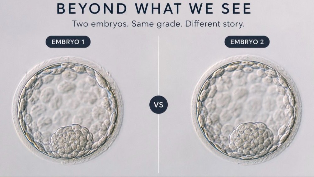
“Two Embryos. Same Grade. Different Story.
Imagine you’re in an IVF laboratory, looking at two blastocysts.
- Both are graded 4AA.
- Both are beautifully expanded.
- Both have a well-defined Inner Cell Mass (ICM) and cohesive Trophectoderm (TE).
If morphology were the only criterion, you might consider them equally promising.
But what if one embryo had a larger ICM, a more uniform TE, a thinner zona pellucida, or more symmetrical blastomeres during its earlier stages of development?
These subtle differences may not be obvious to the naked eye but they can be measured.
This is where morphometric analysis comes into play.
Unlike conventional morphology, which relies on visual assessment, morphometric analysis uses digital image analysis to convert embryo characteristics into objective, measurable data.
Instead of simply describing an embryo as “good” or “well expanded,” it asks questions like:
- How large is the ICM?
- What is the exact blastocyst diameter?
- How symmetrical are the blastomeres?
- How thick is the zona pellucida?
- What is the distance between the pronuclei?
- How much fragmentation is present?
These measurements can be assessed across embryo development.
At the oocyte stage, parameters such as oocyte diameter and zona pellucida thickness can be evaluated.
At the zygote stage, it measures pronuclear size, alignment, distance between pronuclei, and the distribution of nucleolar precursor bodies (NPBs).
At the blastocyst stage, it quantifies blastocyst diameter, blastocoel volume, ICM area, and TE area.
-Why does this matter?
While embryo grading remains the cornerstone of embryo assessment, it is based on visual interpretation.
Morphometric analysis adds objective, reproducible measurements that can reduce observer variability and improve consistency.
With advances in time-lapse imaging and artificial intelligence (AI), these measurements are becoming even more valuable, revealing subtle image-derived features beyond what the human eye can consistently quantify.
Embryo implantation depends on much more than appearance alone. Embryo genetics, endometrial receptivity, maternal factors, and laboratory conditions all influence clinical outcomes.
That’s why morphometric analysis should be viewed as a complementary tool not a replacement for embryologist expertise.
A thought to leave you with…
For decades, we’ve asked:
“Which embryo looks better?”
Perhaps the next question should be:
“What can we learn by measuring what we already see?”
The future of embryo assessment may lie in combining morphology, morphometric analysis, morphokinetics, AI, and the expertise of embryologists to make embryo selection more objective and data-driven.
If two embryos receive the same morphological grade but have different morphometric profiles, should those measurements influence embryo selection? What are your thoughts?”
Stay updated on all scientific advances in the field of fertility with Fertility News.
-
Jun 28, 2026, 17:38Nicole McPherson: New Evidence Suggests Many 1PN Embryos Can Lead to Healthy Live Births
-
Jun 28, 2026, 17:34Yanhe Liu: Study Finds Delayed ICSI Does Not Reduce Cumulative Live Birth Rates in IVF
-
Jun 28, 2026, 17:31Kashif Munir: IVF Lab Contamination Remains a Critical Challenge for Embryo Development
-
Jun 28, 2026, 16:17Madhur Hamine: IVF Is Not Medical Negligence, It Is a National Emergency
-
Jun 28, 2026, 16:14Read Our Special Collection on PPH From FIGO World Congress 2025 – IJGO
-
Jun 28, 2026, 16:10A Machine Learning Model to Predict The Risk of CIN2+ – IJGC
-
Jun 28, 2026, 16:04Adnexal STK11 Tumor Ultrasound Features Featured in New UOGJournal Video – ISUOG
-
Jun 28, 2026, 15:56Paul Pirtea: Should All Oocytes Be Frozen?
-
Jun 28, 2026, 15:37Shaun Greenaway: What People Think Trying for a Baby Looks Like, and What It Actually Feels Like?