

Christopher Robinson: 3D Ultrasound for Delineating Focal Placenta Accreta Spectrum Lesions
Christopher Robinson, Associate Professor Maternal Fetal Medicine at University of South Carolina, shared a post on LinkedIn:
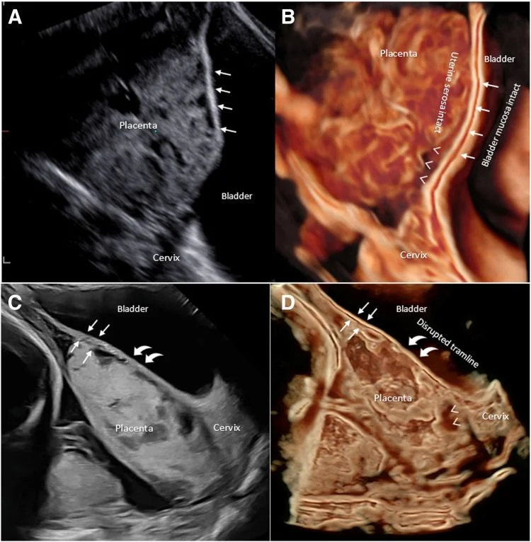
“3-dimensional ultrasound imaging to delineate focal lesions of placenta accreta spectrum – 3D ultrasound tramline sign:
(A) 2D grayscale transvaginal ultrasound (TVS) with straight arrows showing the echogenic line denoting the uterine-bladder interface and (B) in the same patient, 3D TVS rendering (Samsung Crystal–Realistic Vue software), showing 2 continuous echogenic lines (the tramline sign) of the intact uterine serosa and bladder wall (straight arrows).
This image also demonstrates the retroplacental space (arrow heads) along the vesico-uterine interface, extending to the cervix in a case of placenta previa without PAS. (C) 2D grayscale transabdominal ultrasound (TAS) showing the normal vesico-uterine interface (straight arrows), the focally disrupted interface between the uterine serosa and bladder (curved arrows) (D) in the same patient, 3D TAS rendering (GE Healthcare Voluson HD Live mode) showing intact tramline (straight arrows) and a disrupted lower myometrial-placental tramline sign (curved arrows) with absence of the retroplacental space characteristic of focal placenta increta and small area of retroplacental space (arrow heads) present down to the cervix.”
Title: 3-dimensional ultrasound imaging to delineate focal lesions of placenta accreta spectrum
Authors: Savitree Pranpanus, Christoph C. Lees
Read the full article.

Stay updated on all scientific advances in the field of fertility with Fertility News.
-
Jun 19, 2026, 15:54Horace Roman: Sciatic Nerve Endometriosis – Prevent Muscle Atrophy Caused by Progressive Denervation
-
Jun 19, 2026, 15:36Why Does the Clock Read Older? – Fertility Plus
-
Jun 19, 2026, 15:28Asma Khalil: Study Raises Questions About Workplace Activities and Early Pregnancy Outcomes
-
Jun 19, 2026, 15:21Spindle Dynamics and Chromosome Segregation In Human Preimplantation Embryos – Fertility and Sterility
-
Jun 19, 2026, 15:12Today is World Sickle Cell Day – Preeclampsia Foundation
-
Jun 19, 2026, 14:59Marco Zaccaria: ESGE Webinar to Spotlight Advances in the Surgical Management of Parametrial Endometriosis
-
Jun 19, 2026, 14:46PMOS Does Not Make Conception Impossible – PCOS Awareness Association
-
Jun 19, 2026, 14:40Diana Kayal: Successful Fertility Programs Aren’t Built On Clinical Expertise Alone
-
Jun 19, 2026, 14:29Devyanshi Dixit: Call for Abstracts at GFWH 2026