

Ghyass Jutt: Can You Identify a Euploid Blastocyst Under the Inverted Microscope?
Ghyass Jutt, Embryologist at Sakina International Hospital at the Andrology Institute of Pakistan (STAR ICSI), shared on LinkedIn:
“Can you identify a Euploid Blastocyst under the inverted microscope?
The short answer: Not definitively.
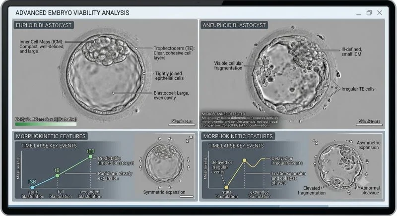
While high-grade morphology and tight cellular cohesion statistically correlate with normal chromosomal status, visual assessment is a proxy, not a diagnostic tool. A visually perfect 5AA blastocyst can be aneuploid, whereas a lower-grade CC embryo can result in a healthy euploid live birth.
Morphological and Morphokinetic Trends
When grading and prioritizing Day 5 or Day 6 blastocysts, embryologists evaluate several key statistical markers:
Euploid Indicators:
- Inner Cell Mass (ICM): Compact, prominent, and densely packed with uniform cells.
- Trophectoderm (TE): Cohesive epithelium with clear, tightly joined cells forming a continuous wall.
- Dynamics: Steady, symmetric blastocoel cavity expansion with on-time developmental milestones.
Aneuploid Red Flags:
- Structural Degeneration: Small or fragmented ICM alongside sparse, uneven TE cells.
- Developmental Delays: Prolonged time to blastulation (delayed into Day 6 or Day 7).
- Morphokinetic Perturbations: Erratic cleavage events, elevated fragmentation, or repeated collapse and re-expansion cycles.
Why Optical Microscopy Reaches Its Limit:
- Mosaicism: Visual inspection cannot resolve cell-level chromosomal variations across trophectoderm and ICM lineages.
- Normal Mitotic Function in Aneuploidy: Aneuploid cells frequently undergo normal mitosis and fluid pumping, creating visually top-grade blastocysts.
- Embryonic Self-Correction: Blastocysts can sequester or extrude aneuploid cells into the perivitelline space while leaving a normal inner cell mass.
High-power light microscopy remains indispensable for embryo selection and prioritization, but trophectoderm biopsy with PGT-A (NGS) remains the sole diagnostic standard for verifying chromosomal status.
How heavily does your laboratory weigh Day 5 vs. Day 6 expansion kinetics when prioritizing transfers in non-PGT-A cycles? Let’s share insights below!”
Stay informed with the latest updates in fertility and reproductive medicine on Fertility News.
-
Sep 20, 2026, 12:44Justin Houman: Honored to Be Selected for Inclusion in 2027 Southern California Super Doctors Rising Stars
-
Sep 20, 2026, 12:41Lorene Lteif: Honored to Have Been a Speaker at the 2nd Trust Fertility and Gynecology Conference 2026
-
Sep 20, 2026, 12:39Maria Satchi: Presenting 2 Topics at BFS Study Week on Male Fertility
-
Sep 20, 2026, 12:36The Most Important Name Tag in Your IVF Journey – Indira IVF Group
-
Sep 20, 2026, 12:34Andris Ābele: Privileged to Participate in the 14th European Congress of Andrology
-
Sep 20, 2026, 12:29The FIGO 2027 Submission Platform Is Open
-
Sep 20, 2026, 11:59Managing Thrombotic Risk in Pregnancy: How the Lab May Help Perspective
-
Sep 20, 2026, 11:20Beyond the Progesterone Number: Rethinking Embryo-Endometrial Synchrony in Frozen Embryo Transfer
-
Sep 20, 2026, 09:49Tommaso Simoncini: Support Not Stigma – 8th World Gynecologic Oncology Day