

Christopher Robinson: AJOG Expert Review Highlights Spiral Artery Transformation Failures in Preeclampsia
Christopher Robinson, Physician/Partner at Charleston Maternal Fetal Medicine, shared a post on LinkedIn about a paper by Eunjung Jung et al. published in AJOG:
“AJOG Expert Review in Preeclampsia: The etiology of preeclampsia – Physiologic transformation of the spiral arteries, failure of physiologic transformation, and atherosis
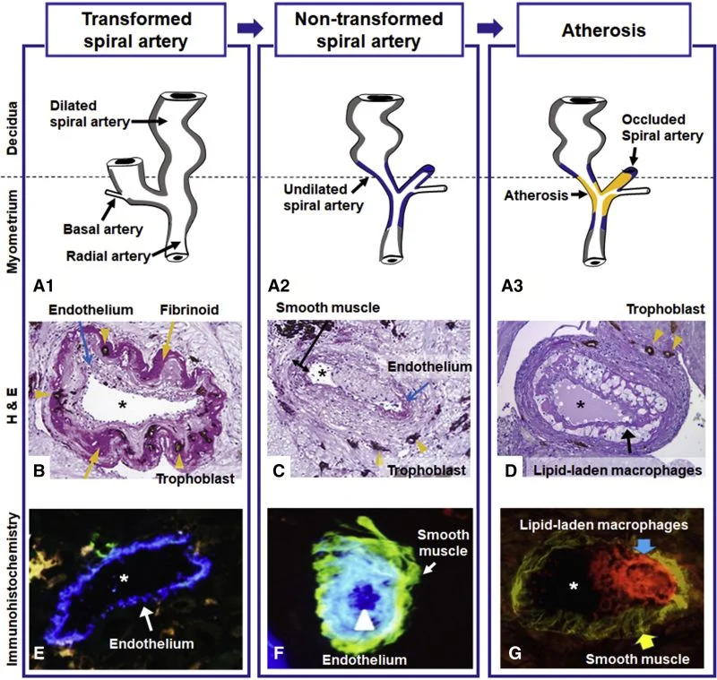
A, Diagram of the maternal blood supply to the placenta. The spiral arteries undergo physiologic changes in normal pregnancy (gray). In preeclampsia, the myometrial segment of the spiral artery fails to undergo physiologic transformation (blue), which is thought to explain the uteroplacental ischemia observed in preeclampsia. Nontransformed spiral arteries are prone to atherosis (yellow), characterized by the presence of lipid-laden macrophages within the lumen. Placental basal plate spiral arteries with hematoxylin-eosin stain (B, C, and D).
B, Transformed spiral arteries are characterized by the presence of intramural trophoblasts (arrowheads) and fibrinoid degeneration (arrows) of the arterial wall.
C, Nontransformed spiral arteries lack intramural trophoblasts and fibrinoid degeneration, and retain smooth muscle. Arrowheads indicate the presence of trophoblasts in myometrium, but not in the wall of the spiral artery.
D, Acute atherosis in a decidual spiral artery. Many lipid-laden macrophages (arrows) are seen in the spiral artery with the lack of invasion of the trophoblast (arrowhead) into a myometrial segment of the spiral artery. Images (B, C, and D) stained with cytokeratin 7 (brown) and periodic acid–Schiff (pink), original magnification ×200. Immunohistochemistry of placental basal plate spiral arteries (E, F, and G).
E, Endothelium (arrow, blue) in vessels with normal trophoblastic invasion, original magnification ×640.
F, A non-transformed spiral artery with endothelium (blue, arrowhead) and smooth muscle cells (green, arrow), original magnification ×640. G, Atherosis lesions show numerous CD36-positive macrophages (red, blue arrow) and smooth muscle cells in the vessel wall (green, yellow arrow), original magnification ×400. Asterisk represents lumen of spiral artery.”
Title: The etiology of preeclampsia
Authors: Eunjung Jung, Roberto Romero, Lami Yeo, Nardhy Gomez-Lopez, Piya Chaemsaithong, Adithep Jaovisidha, Francesca Gotsch, Offer Erez
Read the full article.
Stay updated on all scientific advances in the field of fertility with Fertility News.
-
Oct 11, 2025, 06:44The Global IVF Market Is Set to Reach $65B by 2032 – Meddilink
-
Mar 16, 2026, 13:30Simon Meagher and Rabih Chaoui Discuss Fetal Heart Scanning in the Latest ISUOG Podcast – ISUOG
-
Mar 16, 2026, 12:58Exploring the Link Between Adenomyosis and Proliferative Endometrial Disorders – Fertility and Sterility
-
Mar 16, 2026, 12:55Nicola Pozzi: A New Mechanism to Reduce Thrombus Formation Without Increasing Bleeding Risk
-
Mar 16, 2026, 12:44Endometriosis Awareness Month Virtual Issue! – ISUOG
-
Mar 16, 2026, 12:30Jeanne Conry: When the Government Intervenes, Women Die
-
Mar 16, 2026, 12:23Magdalena Simonis Highlights the Need to Change the Rhetoric on Endometriosis
-
Mar 16, 2026, 12:00Julia Chain Highlights Fertility Preservation as Part of Women’s Health Campaign – HFEA
-
Mar 15, 2026, 15:26Watching Life Begin: Scientists Track Embryo Implantation in Real Time! – Science Magazine
-
Mar 15, 2026, 15:03Reem Abu-Rustum Spotlights Fetal Situs in New ISUOG Quiz – ISUOG