

Somil Singhal: Embryo Selection – Map To Testing
Somil Singhal, Medical Doctor at AIIMS (All India Institute of Medical Sciences, New Delhi), shared on LinkedIn:
“Embryo Selection: Map To Testing
New technologies are making extraordinary progress in fertility diagnostics.
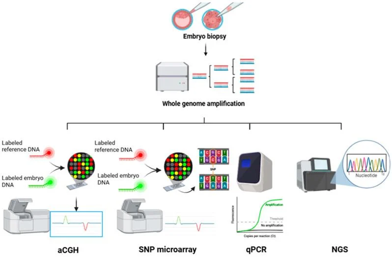
One of the latest advances incorporated into the range of diagnostic techniques is called array-CGH.
This genetic test allows to analyze cells’ DNA to limits.
The human genome is made up of 46 chromosomes: 23 from the father and 23 from the mother.
All of our organism’s genetic information is stored in these chromosomes.
The excess or defect of any chromosome region may be responsible for certain diseases or fertility problems, such as implantation failure or recurrent miscarriages.
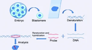
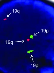
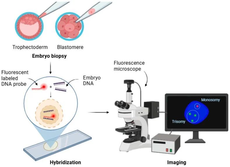
FISH: the use of fluorescent probes allows to analyze the presence or absence of a single chromosomal region.
The Array-CGH is much more sensitive and efficient than the conventional karyotype and allows to reach diagnostic levels that are 10 times higher than the karyotype.
Karyotype is the ‘general map’ of chromosomes in which one can see only the highest mountains and the largest rivers, whereas the Array-CGH would act like a zoom that would greatly expand the resolution of map so that the smaller mountains and rivers and would become visible.
This means that the chromosomal analysis through Array-CGH allows to detect chromosomal abnormalities that would be imperceptible in the karyotype.
The Array CGH technique analyzes the complete genome of an individual looking for gains or losses of genetic material.
Array-CGH allows to identify duplications or absences of small chromosomal regions that the karyotype is not able to detect.
With an Array-CGH analysis, the DNA problem is marked in fluorescent green.
In parallel, a DNA control (without any chromosomal abnormality) is marked in red.
The mixture of the two fluorescent labels is hybridized against a DNA “chip”.
This chip or array contains a collection of molecules of DNA that sweep the entire human genome.
Applications of this technique:
Preimplantation diagnosis:
To select embryos that did not implant or caused a miscarriage in an IVF cycle due to alterations in the chromosomes.
Prenatal diagnosis:
To identify possible chromosomal alterations in foetuses in which anomalies were detected by ultrasound.
Complete molecular karyotype for couples with fertility problems due to unknown causes.
Array-CGH is a highly promising diagnostic and therapeutic tool, since it allows to identify chromosomal alterations that could not be detected by other techniques.
It therefore offers a solution to many couples that come in their search for a healthy baby of their own.
This advanced diagnostic tool represents a major leap forward from traditional chromosome analysis.
By scanning the fetal genome at an incredibly high resolution, it allows to detect chromosomal imbalances that would otherwise go unnoticed, helping families and healthcare providers make informed management decisions.
Array CGH Performed During Pregnancy.
Array CGH is not a routine screening test.
Instead, it is recommended under specific circumstances, including:
- Abnormal Ultrasound Findings: Detection of structural anomalies in the fetal heart, brain, kidneys, or skeletal system, or severe unexplained intrauterine growth restriction (IUGR).
- High-Risk Prenatal Screening Results: Concerning findings from maternal serum screening or non-invasive prenatal testing that suggest a chromosomal abnormality.
- Family History of Genetic Conditions: A previous child or family member with unexplained intellectual disability, developmental delay, or multiple congenital anomalies.
- Recurrent Pregnancy Loss: To investigate whether chromosomal imbalances are contributing to recurrent miscarriages.
To perform Array CGH, actual fetal cells must be obtained.
This is done through one of two invasive diagnostic procedures:
- Chorionic Villus Sampling (CVS): Typically performed between weeks 11 and 14 of pregnancy, where a small sample of placental tissue is collected.
- Amniocentesis: Typically performed after week 15 of pregnancy, where a small amount of amniotic fluid surrounding the fetus is drawn using a fine needle under ultrasound guidance.
Understanding the Results and the Role of Genetic Counseling.
Once the sample is processed, results fall into three categories:
Normal Result: No clinically significant copy number variants (gains or losses) were detected. While highly reassuring, it does not rule out single-gene disorders or conditions below the test’s resolution.
Pathogenic Result: A deletion or duplication associated with a known genetic syndrome or developmental disorder was identified, helping the medical team plan appropriate care.
Variant of Uncertain Significance (VUS): A genetic alteration was found, but its clinical impact is currently unknown.
In such cases, testing the biological parents is often recommended to determine if the variant is inherited (usually benign) or occurred spontaneously.”
Proceed to the video attached to the post.
Other articles featuring Somil Singhal on Fertility News.
-
Oct 24, 2026, 17:12Rana Mondal: Can We Replace Daily Fertility Injections with a Simple Daily Pill?
-
Oct 24, 2026, 17:03Evaluating Hysteroscopic Subendometrial Human Umbilical Cord Blood Platelet-Rich Plasma Injection – RBMO
-
Oct 24, 2026, 16:56Muhammad Qasim: Excluded Cells and Embryo Biopsy
-
Oct 24, 2026, 16:48Sangita Jindal: Completing My Term on the ASRM Board of Directors as Chair of the Publications Committee
-
Oct 24, 2026, 16:33Clare Mansfield: Flowering Fertility Support Programme Was Independently Evaluated by Health Innovation Hub Ireland
-
Oct 24, 2026, 16:17Diana Tain: Vitrification May Affect Oocyte Resilience Without Causing Chromosomal Instability
-
Oct 24, 2026, 16:02Albert Irkaliev: Peptides and the Future of ART
-
Oct 24, 2026, 15:57Resa Nabor: A Filipino Woman’s Journey of Faith, Infertility, and Unwavering Love
-
Oct 24, 2026, 15:41Esther Eleboh: Comparison of IVF Ovarian Stimulation Protocols