

Shivam Jha: What is the Importance of Ultrasound During Pregnancy?
Shivam Jha, Clinical Application Specialist – North at Philips, shared a post on LinkedIn:
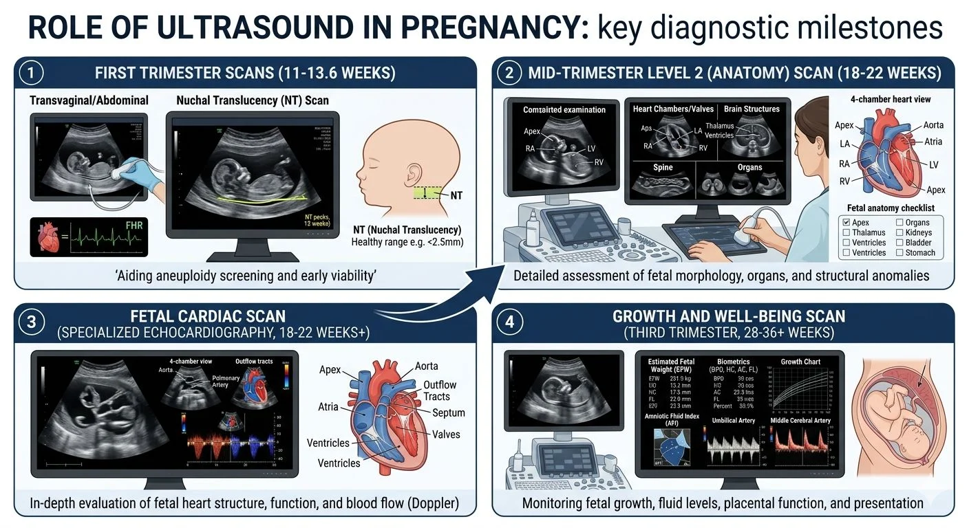
“What is the Importance of Ultrasound During Pregnancy?
1. Cardiac Activity Scan
A cardiac activity scan, typically conducted between 6-7 weeks of pregnancy, provides information about the duration of pregnancy by the length of the embryo – CRL. It also confirms whether the pregnancy is viable and live by detecting the presence and strength of a developing heartbeat.
2. First Trimester Scan
NT NB ultrasound or Level 1 ultrasounds look for Nuchal translucency, Nasal Bone with other structural and doppler parameters to ascertain with a Dual marker test that the developing fetus is healthy and doesn’t have any genetic syndrome like Down syndrome. Usually performed between 11 weeks to 14 weeks of pregnancy.
3. Level 2 Scan
A Level 2 ultrasound is performed between 18 to 22 weeks of pregnancy. Level 2 ultrasound is performed to produce images of the fetus and its internal organs. The ultrasound helps to assess the size and growth of the foetus, evaluate the formation of the brain, face, spine, heart, lungs, and other organs, and to identify any potential structural abnormalities.
In addition, the ultrasound can help determine the placenta’s position, evaluate the amount of amniotic fluid surrounding the fetus, and assess the blood flow in the umbilical cord.
3D ultrasound (and even 4D) is becoming more common and popular.
4. Fetal Echo Scan
A fetal echo scan is an ultrasound test used to examine the heart of a developing fetus. Fetal echo scans are typically performed between 19 and 22 weeks of pregnancy and are commonly used to diagnose heart problems in the fetus.
Fetal echo scans are important because early detection and treatment of heart problems in the fetus can significantly improve outcomes for both the mother and the baby.
Doppler and Growth Scans
A growth scan is typically conducted in the third trimester of pregnancy to evaluate the developing fetus’s growth and well-being. This scan assesses various parameters such as the fetal size, weight, and the amount of amniotic fluid present in the womb. Generally, the first growth scan is done at around 28 weeks, and further scans, if needed, are decided based on the first scan’s findings. If everything appears normal, a second growth scan is typically performed around 36 weeks, which is often combined with a Doppler scan.
A Doppler scan measures the blood flow through the umbilical cord and different parts of the fetal body, such as the fetal brain and liver. This scan can indicate whether the fetus receives enough oxygen and nutrients through the placenta.
Conclusion!
Ultrasound plays a crucial role in pregnancy, providing valuable information about the growth and development of the fetus, placenta, and surrounding structures. Whether it is transabdominal ultrasound or 3-D, from observing blood flow and detecting abnormalities, ultrasound is a powerful tool for expectant parents and healthcare providers to produce detailed images of the baby.”
Stay updated on all scientific advances in the field of fertility with Fertility News.
-
Apr 1, 2026, 13:32Pregnancy Loss Leave and Recognition in Ireland! – PLRG
-
Apr 1, 2026, 13:19Christopher Robinson: Immune Tolerance Breakdown In Pregnancy!
-
Apr 1, 2026, 13:14How Many Cells Are Enough in Blastocyst Biopsy? – Fertility Plus
-
Apr 1, 2026, 13:09Follicular Fluid PFAS May Influence IVF Outcomes – Fertility and Sterility
-
Apr 1, 2026, 12:48Taarika Ramesh: Rethinking IVF Stimulation With Progestin-Primed Protocols
-
Apr 1, 2026, 12:18Abnormal Spermogram Is Not an Explanation, It Is a Starting Point – Cesar Fertility
-
Apr 1, 2026, 11:41Christopher Robinson: Racial Disparities in Postoperative Benign Hysterectomy Outcomes
-
Apr 1, 2026, 11:26Sergei Gorlovetsky: Most Fertility Clinics Are Losing Patients Between Interested and Started Treatment
-
Apr 1, 2026, 11:16CFEF Launches Secure Forum for Fetal Ultrasound Professionals – CFEF