

Christopher Robinson: AJOG Expert Review Highlights Spiral Artery Transformation Failures in Preeclampsia
Christopher Robinson, Physician/Partner at Charleston Maternal Fetal Medicine, shared a post on LinkedIn about a paper by Eunjung Jung et al. published in AJOG:
“AJOG Expert Review in Preeclampsia: The etiology of preeclampsia – Physiologic transformation of the spiral arteries, failure of physiologic transformation, and atherosis
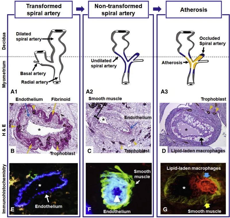
A, Diagram of the maternal blood supply to the placenta. The spiral arteries undergo physiologic changes in normal pregnancy (gray). In preeclampsia, the myometrial segment of the spiral artery fails to undergo physiologic transformation (blue), which is thought to explain the uteroplacental ischemia observed in preeclampsia. Nontransformed spiral arteries are prone to atherosis (yellow), characterized by the presence of lipid-laden macrophages within the lumen. Placental basal plate spiral arteries with hematoxylin-eosin stain (B, C, and D).
B, Transformed spiral arteries are characterized by the presence of intramural trophoblasts (arrowheads) and fibrinoid degeneration (arrows) of the arterial wall.
C, Nontransformed spiral arteries lack intramural trophoblasts and fibrinoid degeneration, and retain smooth muscle. Arrowheads indicate the presence of trophoblasts in myometrium, but not in the wall of the spiral artery.
D, Acute atherosis in a decidual spiral artery. Many lipid-laden macrophages (arrows) are seen in the spiral artery with the lack of invasion of the trophoblast (arrowhead) into a myometrial segment of the spiral artery. Images (B, C, and D) stained with cytokeratin 7 (brown) and periodic acid–Schiff (pink), original magnification ×200. Immunohistochemistry of placental basal plate spiral arteries (E, F, and G).
E, Endothelium (arrow, blue) in vessels with normal trophoblastic invasion, original magnification ×640.
F, A non-transformed spiral artery with endothelium (blue, arrowhead) and smooth muscle cells (green, arrow), original magnification ×640. G, Atherosis lesions show numerous CD36-positive macrophages (red, blue arrow) and smooth muscle cells in the vessel wall (green, yellow arrow), original magnification ×400. Asterisk represents lumen of spiral artery.”
Title: The etiology of preeclampsia
Authors: Eunjung Jung, Roberto Romero, Lami Yeo, Nardhy Gomez-Lopez, Piya Chaemsaithong, Adithep Jaovisidha, Francesca Gotsch, Offer Erez
Read the full article.
Stay updated on all scientific advances in the field of fertility with Fertility News.
-
Jul 29, 2026, 01:03Seeking a Second Opinion Is Not Being Difficult, It Is a Valid Part of Informed Care – PCOS Awareness Association
-
Jul 29, 2026, 00:57Szabo Gabor: IOTA Day 2026 Advancing Ultrasound in Gynaecological Oncology
-
Jul 29, 2026, 00:55Islam Badr: A Simple Approach to Fetal Heart Screening
-
Jul 29, 2026, 00:50Hardeep Singh: Understanding the Global IVF Travel Landscape
-
Jul 29, 2026, 00:48Asma Khatun: Shaping the Future of Women’s Healthcare at GFWH 2026
-
Jul 29, 2026, 00:43Shaping the Future of Reproductive Medicine at ASRM Innovate – ASRM
-
Jul 29, 2026, 00:37George Condous: Gratitude Makes Every Achievement Greater!
-
Jul 29, 2026, 00:35As AMH Falls, Progesterone May Rise Earlier in Ovarian Stimulation! – Fertility Plus
-
Jul 29, 2026, 00:33An Easy Method for Systematic Fetal Cardiac Screening – ISUOG