

Alisha Tuck: Understanding Endometrial Carcinoma on Ultrasound
Alisha Tuck, OB Gyn, Sonographer at Sonopartners LLC, shared a post on LinkedIn:
“Understanding Endometrial Carcinoma on Ultrasound
Endometrial carcinoma is the most common gynecologic malignancy in developed countries, primarily affecting postmenopausal women. While a definitive diagnosis requires a tissue biopsy (usually via dilation and curettage or office pipelle), Transvaginal Ultrasound (TVUS) serves as the primary frontline imaging tool for screening and staging.
1. Clinical Presentation and Risk Factors
The hallmark symptom of endometrial cancer is postmenopausal bleeding (PMB). In premenopausal patients, it may manifest as heavy, prolonged, or irregular menstrual cycles.
Key Risk Factors:
- Unopposed Estrogen: Obesity (where peripheral fat converts precursors to estrogen), estrogen-only hormone replacement therapy, or PCOS.
- Tamoxifen Use: Often used for breast cancer treatment, it can have a pro-estrogenic effect on the uterus.
- Lynch Syndrome: A genetic predisposition to various cancers.
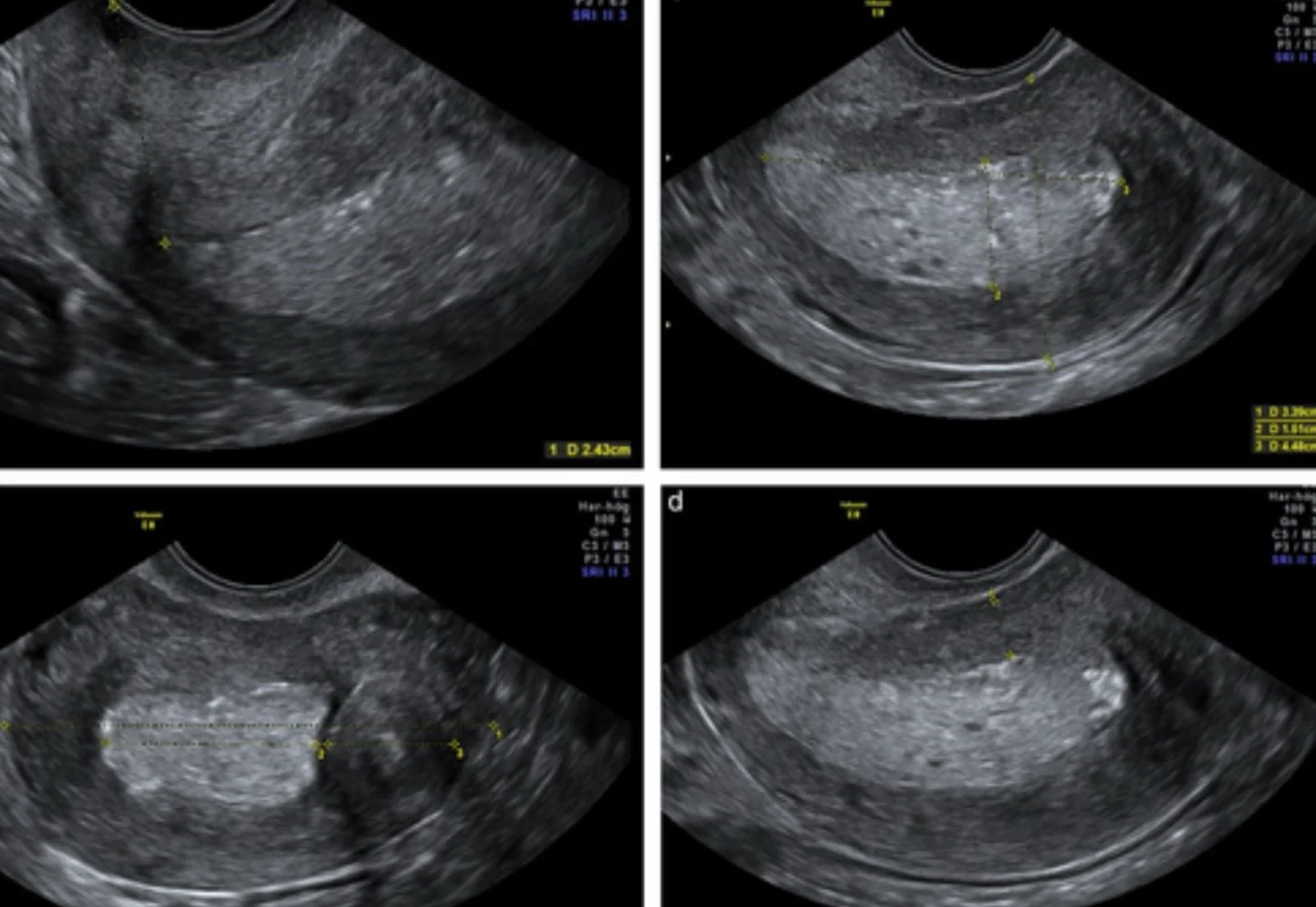
2. Sonographic Hallmarks
When evaluating the uterus for malignancy, sonographers and radiologists look for several specific “red flags.”
- Endometrial Thickness (ET): In postmenopausal women with bleeding, an ET > 4 mm is generally considered the threshold for further investigation.
- In asymptomatic postmenopausal women, the threshold is higher (often > 8–11 mm), though this is debated.
- Echogenicity: The endometrium often appears heterogeneous, thickened, and “dirty” or complex compared to the surrounding tissue.
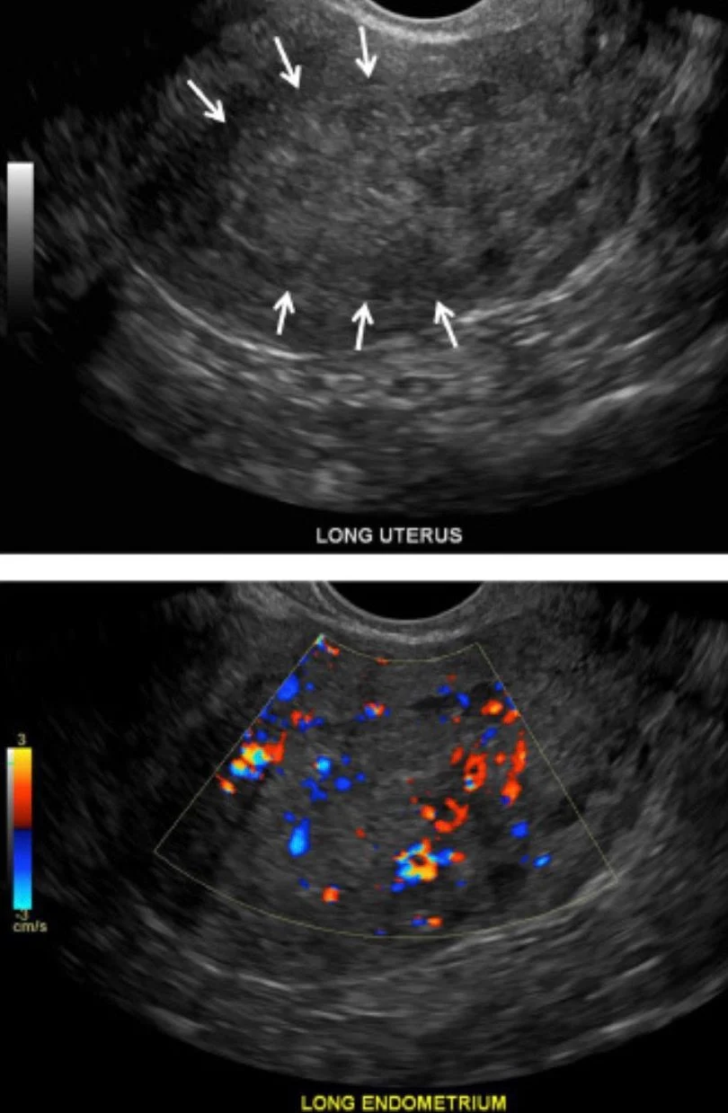
- The Subendometrial Halo: A healthy uterus has a thin, hypoechoic (dark) zone between the endometrium and myocardium. Loss or blurring of this ‘halo’ suggests myometrial invasion.
- Fluid Collections: While simple fluid (hydrometra) can be benign, fluid containing echoes (hemometra or pyometra) in a postmenopausal patient is suspicious.
3. The Role of Doppler Imaging
Color and Power Doppler are essential for differentiating between benign polyps and malignant masses.
- Neovascularization: Malignant tumors often display ‘multiple vessel’ patterns with irregular branching.
- Resistive Index (RI): Malignant flow often exhibits low resistance.
The primary goal of the ultrasound, beyond detection, is to determine how deep the cancer has grown into the uterine wall. This is critical for surgical planning.
Note: MRI is often superior to ultrasound for precise staging (especially for cervical involvement), but TVUS remains the most cost-effective initial assessment.
4. Limitations of Ultrasound
It is important to remember that ultrasound cannot always distinguish between:
- Endometrial Polyps: Often more focal and have a single ‘feeding vessel.’
- Endometrial Hyperplasia: A precursor to cancer that can look identical to early-stage carcinoma.
- Submucosal Fibroids: These can distort the endometrial lining but usually have a distinct, striated appearance.
Disclaimer: For educational purposes only. Correlate with clinical symptoms and histopathological results.”
Stay updated on all scientific advances in the field of fertility with Fertility News.
-
Jul 1, 2026, 19:47Marian Knight: Fantastic Day at the Oxford Women’s Health Forum
-
Jul 1, 2026, 19:36Rachel Small: Early Pregnancy Care Must Not Be Left Out of Maternal Health Discussions
-
Jul 1, 2026, 19:19Why Has Infertility Been Increasing in Recent Years? – MEFS
-
Jul 1, 2026, 19:10Francesca Crovetto: Can Pregnancy Complications Be Prevented Before Glucose Becomes Abnormal?
-
Jul 1, 2026, 18:54Aumatma Simmons: Clinical Implications for Reproductive Health – Circadian Rhythm Optimization
-
Jul 1, 2026, 16:01ASRM Launches Educational Podcast Series to Support LGBTQ+ Family-Building Journeys
-
Jul 1, 2026, 15:40Vakkanal Paily Awarded RCOG Distinguished Service Medal for Transforming Maternal Care in Kerala – RCOG
-
Jul 1, 2026, 15:11It All Comes Down to One Molecule – VEGF – Fertility Plus
-
Jul 1, 2026, 12:02Christopher Robinson: Impact of Maternal Aspirin Therapy on Neonatal Epigenetic Patterns