

Awais Hamza: Neural Tube Defects – Embryology Meets Prenatal Ultrasound
Awais Hamza, Virtual Medical Assistant at SNS Rheumatology Associates, shared a post on LinkedIn:
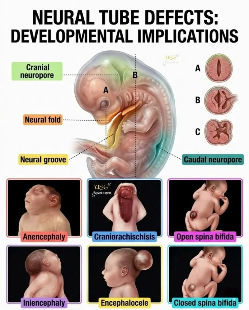
“Neural Tube Defects (NTDs): Embryology Meets Prenatal Ultrasound
Neural tube defects occur when the neural tube fails to close properly during the first 4 weeks of embryonic development. The location and timing of closure failure determine the type and severity of the anomaly.
Early prenatal ultrasound plays a critical role in detecting these conditions, counseling families, and guiding pregnancy management.
Key Neural Tube Defects & Ultrasound Findings
Anencephaly • Failure of cranial neuropore closure • Absence of calvarium and cerebral hemispheres • Classic ultrasound appearance: ‘Frog-eye sign’ • Detectable as early as the first trimester • Lethal condition
Craniorachischisis • Failure of both cranial and spinal neural tube closure • Combination of anencephaly with extensive open spinal defect • Ultrasound: absent cranial vault with exposed neural tissue extending along the spine • Rare but severe neural tube defect
Open Spina Bifida (Myelomeningocele) • Failure of caudal neuropore closure • Ultrasound findings:
Spinal defect with protruding sac
Lemon sign (frontal bone scalloping)
Banana sign (curved cerebellum)
Ventriculomegaly may be present • Often associated with Chiari II malformation
Closed Spina Bifida • Skin covers the defect • More subtle prenatal sonographic findings • May show soft tissue mass, lipoma, or vertebral abnormalities • Neurological impairment varies
Encephalocele • Herniation of intracranial contents through a skull defect • Usually occipital in location • Ultrasound: extracranial cystic or solid mass connected to the cranial vault • Evaluate contents carefully (meninges vs brain tissue)
Iniencephaly • Severe cervical spine malformation with extreme retroflexion of the fetal head • Short spine and defective occipital bone • Ultrasound: fixed hyperextension of the head and neck • Often associated with other congenital anomalies
Sonographer’s Pearl Whenever an open neural tube defect is suspected, carefully evaluate:
- Posterior fossa
- Ventricular system
- Spine in sagittal, transverse, and coronal planes
- Associated intracranial markers
- Amniotic fluid volume and associated anomalies
Early diagnosis allows multidisciplinary counseling and optimized prenatal care.”
Stay updated on all scientific advances in the field of fertility with Fertility News.
-
Jun 21, 2026, 14:10Christopher Robinson: Substance Use and Use Disorders During Pregnancy and the Postpartum Period
-
Jun 21, 2026, 13:48FIGO – WHO Webinar on Updated Medical Eligibility Criteria – IJGO
-
Jun 21, 2026, 13:40Webinar On Abdominal Repair of High Vaginal Tears with Haemodynamic Compromise – GEFOG Health Foundation
-
Jun 21, 2026, 12:13Fereshteh Safian: A Case From the IVF Lab On Dysmorphic Oocytes and Embryo Development
-
Jun 21, 2026, 12:06Evelyn Ndinda: Prolactin as a Key Biomarker in Female Infertility
-
Jun 21, 2026, 11:58Reema Sircar: Fertility Preservation Is Empowering Only With Real Numbers
-
Jun 21, 2026, 11:46Abdul Mannan: Pregnancy Changes Almost Every Coagulation Test
-
Jun 21, 2026, 11:28What Is a Biochemical Pregnancy and Why Does It Happen? – Fertility Plus
-
Jun 21, 2026, 11:24Advocating for Health Equality Through Fibroid Awareness – Fibrome Info France