

Lydia Akpan: Key Signs of Oocyte Dysmorphism
Lydia Akpan, Embryologist Trainee at St. Ives Specialist Hospital, shared a post on LinkedIn:
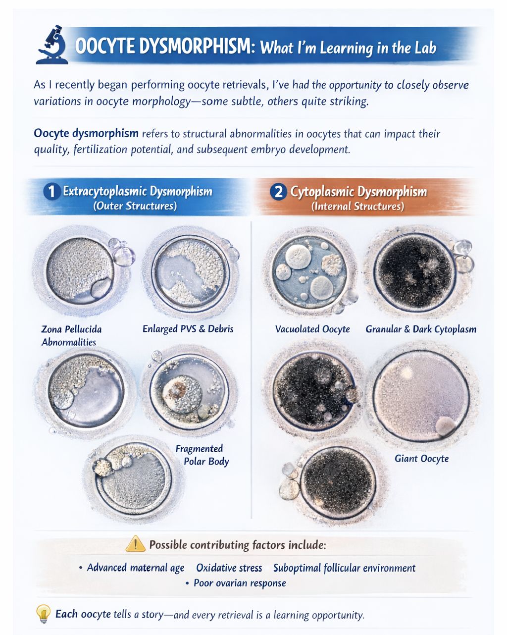
“OOCYTE DYSMORPHISM: What I’m Learning in the Lab
As I recently began performing oocyte retrievals, I’ve had the opportunity to closely observe variations in c—some subtle, others quite striking.
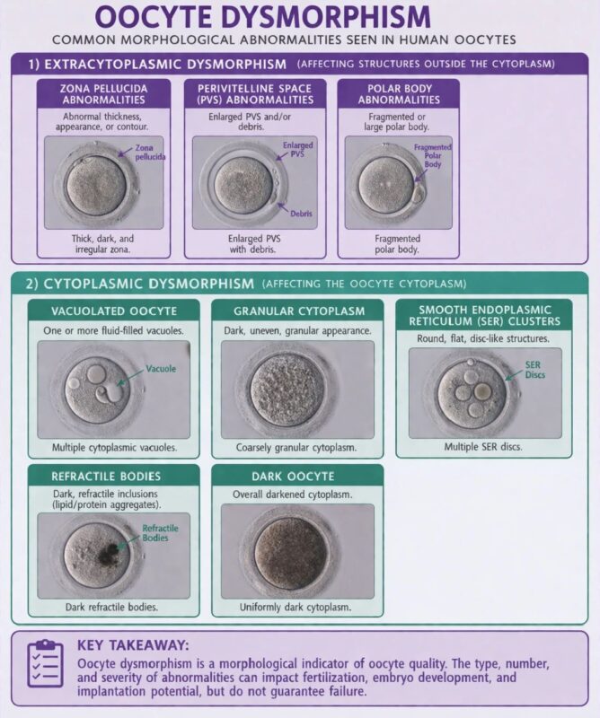
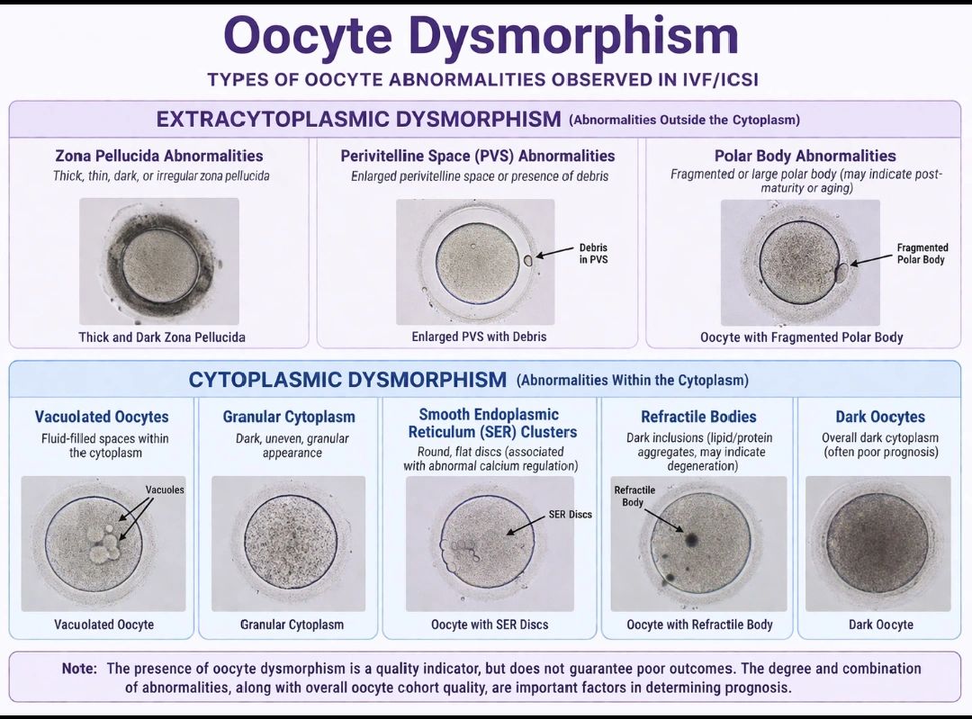
OOCYTE DYSMORPHISM: refers to structural abnormalities in oocytes that can impact their quality, fertilization potential, and subsequent embryo development.
These abnormalities are broadly classified into:
1. Extracytoplasmic Dysmorphism (Outer Structures)
- Zona pellucida abnormalities – Thick, thin, dark, or irregular; may affect sperm binding and fertilization.
- Perivitelline space (PVS) abnormalities – Enlarged space or presence of debris/fragments.
- Polar body abnormalities – Fragmented or enlarged; may indicate post-maturity or aging.
2. Cytoplasmic Dysmorphism (Internal Structures)
- Vacuolated oocytes – Fluid-filled inclusions that may disrupt spindle formation.
- Granular cytoplasm – Dark, uneven appearance linked to poorer embryo quality.
- Smooth endoplasmic reticulum (SER) clusters – Associated with abnormal calcium regulation.
- Giant oocytes – Abnormally large; may contain extra chromosomal material and increase risk of aneuploidy.
- Refractile bodies – Dense inclusions, possibly indicating degeneration.
- Dark oocytes – Often associated with poor prognosis
Possible contributing factors of dysmorphism include:
- Poor ovarian stimulation response
- Advanced maternal age
- Oxidative stress
- Suboptimal follicular environment
- Lab handling conditions.
Each oocyte tells a story—and every retrieval is a learning opportunity.”
Stay updated on all scientific advances in the field of fertility with Fertility News.
-
Jul 25, 2026, 10:56PMOS Can Affect Much More Than Periods or Fertility – PCOS Awareness Association
-
Jul 25, 2026, 10:42Asma Khalil: Recreational Drug Use During Pregnancy, What Every Expectant Mother Should Know!
-
Jul 25, 2026, 10:40Khalil Faaed: Are Complex Y Chromosome Rearrangements the Missing Link in Unexplained Male Infertility?
-
Jul 25, 2026, 10:38Sean Lauber: Does Transferring a Cleavage or Blastocyst Stage Embryo Affect the Chance of Having a Boy?
-
Jul 25, 2026, 10:33Rokaya Hachicho: The Hidden Cost of Excessive Laser Use During Trophectoderm Biopsy!
-
Jul 25, 2026, 10:31Join the ISUOG Webinar on Late-Evolving Fetal Anomalies – ISUOG
-
Jul 25, 2026, 10:29Waiting Before Clamping the Umbilical Cord Improves Neonatal Outcomes! – RCOG
-
Jul 25, 2026, 10:27Christopher Robinson: The Impact of Body Mass Index on Misoprostol Dosing for Labor Induction!
-
Jul 25, 2026, 10:26Heavy Menstrual Bleeding and Dysmenorrhea in Adolescents – IJGO