
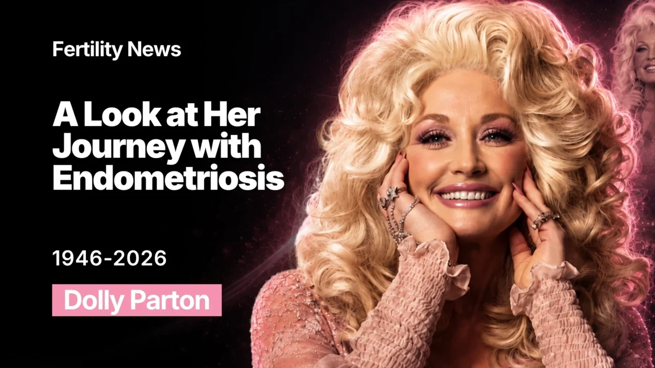
Dolly Parton: A Look at Her Journey With Endometriosis
Dolly as a Country Music Icon
Dolly Parton’s story has long been associated with music, extraordinary longevity in the entertainment industry, and a life without biological children. But behind that decision was also a significant gynecologic history linked to endometriosis.
Dolly Parton (1946–2026) was an American country music icon, singer-songwriter, actress, entrepreneur, and philanthropist, celebrated for transforming personal, Appalachian-rooted storytelling into worldwide popular music and for using her success to support children’s literacy and other charitable causes.
She was born into a large family, the fourth of twelve children, on January 19, 1946, in Locust Ridge, Tennessee, and came from deep in Appalachia, where music was an integral part of life for those who, like the Partons, struggled to make a hard living. Her mother was a singer who taught Dolly both church music and the Elizabethan ballads her ancestors brought to America. Dolly’s grandfather was a fiddling preacher who wrote “Singing His Praise,” which was recorded by Kitty Wells. Several of Dolly’s eleven siblings have been active in music, and some worked for a time in her family band.
Parton’s music career progressed apace; people began to take note of her as a songwriter, especially after a pair of songs she wrote with Owens became Top Ten hits for Bill Phillips in 1966. Then she recorded for Fred Foster’s Monument Records from 1965 to 1967, and “Dumb Blonde” – which attacked traditional female stereotypes – became her first Top Forty hit.
Dolly Parton’s Endometriosis Journey
Dolly Parton’s decision not to become a mother was not simply a personal choice. At a very young age, she was diagnosed with endometriosis. Parton reportedly had to cancel a tour in the early ‘80s due to severe abdominal pain and bleeding – common symptoms of the condition. She underwent a partial hysterectomy in 1984, when she was in her 30s, and was unable to have biological children afterward. Parton later spoke about how profoundly the experience affected her, both physically and emotionally.
“I can’t have children physically. I love children. I don’t know that it also wasn’t God’s will, you know, for me not to have kids. I mean, I try to look at things in a way that I can put them in a proper place because somebody has to be out there doing something to entertain other people’s children,” she said at the time.
Dolly Parton’s openness about her struggle with endometriosis sparked a vital global conversation, empowering countless women to recognize, discuss, and seek treatment for gynecological health issues. Fortunately, medical advancements mean that a partial hysterectomy is no longer the definitive solution. Today, patients with endometriosis have access to a wider variety of advanced, fertility-preserving treatments, offering hope to those who still wish to conceive biological children.

Endometriosis: A Chronic Disease With Reproductive Consequences
Endometriosis is a chronic medical condition where tissue similar to the inner lining of the uterus grows outside of the uterus. It influences 10%-15% of all reproductive-age females and 70% of women with persistent pelvic pain. Endometriosis pathogenesis suggests that the disease’s etiology is complicated and multifaceted, involving genetic, hormonal, immunological, and environmental factors.
The ovaries and pelvic peritoneum are the most common sites for developing endometriotic lesions, which can also develop in the fallopian tubes, bowel, bladder, abdominal wall, cervix, and vagina. The pathophysiology of endometriosis and pain is poorly known, with most gynecologists believing that inflammation is a crucial source of irritation in endometriosis.
Why and How Endometriosis Emerges
The definite pathogenesis of endometriosis is still unknown, but there are a number of leading theories including retrograde menstruation, altered immunity, coelomic metaplasia, and metastatic spread.
1. Retrograde Menstruation
The most well-accepted theory, retrograde menstruation, states that endometrial tissue is transported in a retrograde fashion through patent fallopian tubes into the peritoneal cavity. The endometrial cells then attach to the peritoneal mesothelial cells, establish a blood supply, proliferate, and produce endometrial implants. This theory has been well supported by subsequent research.
Women with endometriosis have higher volumes of refluxed menstrual blood and endometrial-tissue fragments than women without the disorder. Similarly, young girls with obstructed outflow tracts show higher rates of the disease. Still, retrograde menstruation occurs at similar rates in women with and without endometriosis, suggesting other factors must also contribute.
2. Altered Immunity
Women with endometriosis have altered immunity, preventing them from clearing the refluxed endometrial cells/fragments that appear in retrograde menstruation. This would help explain why some women with retrograde menstruation develop endometriosis while others do not. Cell-mediated immunity is thought to be deficient in patients with the disease; leukocytes are unable to recognize that the endometrial tissue is not in its normal location.
In women with endometriosis, there are increased numbers of leukocytes and macrophages in and around endometrial implants and in the peritoneal fluid. These cells secrete cytokines and growth factors (IL-1, 6, and 8, TNF, RANTES, VEGF) into the peritoneal milieu, which then recruit surrounding capillaries and leukocytes. The ultimate effect is proliferation of endometriosis implants with increased vascular supply.
3. Coelomic Metaplasia and Metastatic Spread
In the 1960’s, Ferguson proposed that coelomic metaplasia may also contribute to the development of endometriosis. It stems from the theory that the peritoneum contains undifferentiated cells that can differentiate into endometrial cells.
In addition to retrograde menstruation, coelomic metaplasia, and altered immunity, newer research is increasingly showing that stem cells and genetics may play a role in the etiology of endometriosis.
4. Stem Cells
It is presumed that de novo development of endometrial tissue occurs from endogenous stem cells in the endometrium. Over the last decade, we have studied the possibility that bone marrow-derived cells may also differentiate into endometrial cells, and pertinently, may be implicated in the development of ectopic endometrial implants. If true, this would help explain how ectopic tissue can occur in locations outside the peritoneal cavity such as the lung and CNS system.
Proof that endometrial cells can be derived from bone marrow mesenchymal stem cells comes from the study of female allogeneic bone marrow transplant recipients who received marrow from a single antigen-mismatched related donor, allowing the cells to be identifiable by HLA type.
The study remarkably showed the presence of donor-derived endometrial cells in endometrial biopsies of the recipients. This finding suggested that bone marrow-derived stem cells can differentiate into human uterine endometrium. The endometrial tissue must be capable of attracting stem cells despite its ectopic location. This suggests an alternative origin of some endometriosis, specifically, from bone marrow-derived cells.
5. Genetics
For over 20 years, it has been known that endometriosis has a familial tendency. Women who have a first-degree relative affected by the disease have a 7 times higher risk of developing endometriosis than women who do not have a family history of the disease.
Clinical Presentation of Endometriosis
Clinical presentation of endometriosis varies in women. Patients often present with symptoms such as intermenstrual bleeding, painful periods (dysmenorrhea), painful intercourse (dyspareunia), painful defecation (dyschezia), and painful urination (dysuria). Pelvic pain may present before menstruation begins. Often, endometriosis can be asymptomatic, only coming to a clinician’s attention during evaluation for infertility.
The American Society for Reproductive Medicine (ASRM) classifies endometriosis into four stages based on the extent and location of lesions and adhesions:
- stage I (minimal),
- stage II (mild),
- stage III (moderate),
- stage IV (severe).
Several reproductive factors also affect the risk. Earlier menarche and shorter menstrual cycles are linked to higher risk, while having children and current oral contraceptive use are linked to lower risk. Lower body mass index has also been associated with the condition.
How does endometriosis get treated nowadays?
It is well known that endometriosis is an estrogen-dependent disorder. Endometriotic lesions have been shown to have an increased production and decreased inactivation of estradiol. Common medical therapies used to treat symptoms of endometriosis, such as pelvic pain, dyspareunia, and dysmenorrhea, target ovarian estrogen production.
Medications used as endometriosis therapy are hormonal medications including combined oral contraceptives, progestins, danazol, and gonadotropin-releasing hormone agonists or antagonists (GnRH analogs). Although these medications may help treat pain, they have shown no benefit in the treatment of endometriosis-associated infertility.
The recommended daily dose of danazol for the treatment of endometriosis is 600-800 mg; even so, the dose has significant steroid side effects, including increased hair growth, changes in mood, an irreversible deepening of the voice, bad impacts on serum lipids, and, in rare cases, irreversible and life-threatening liver injury.
Hormone therapy often prevents ovulation by preventing the ovaries from releasing hormones, including estrogen. This could aid in reducing the rate of local development and activity of the endometrium and endometrial lesions. Other medications have been inadequately investigated, including clomiphene, tamoxifen, and the anti-progestational drug mifepristone.
Surgical techniques include excision or removal of endometrial implants, ablation of uterosacral nerves by employment of endocoagulation, electrocautery, or laser treatment, presacral neurectomy, and hysterectomy with bilateral salpingooophorectomy. They have a 50–80% success rate in reducing symptoms. Unfortunately, endometriosis recurs in 5 to 15% of cases even after hysterectomy and bilateral oophorectomy.
Endometriosis pain is treated surgically by interrupting the brain circuits that carry pain signals. Various considerations should be taken, including the best method of gaining access to the pelvis and abdomen, which can be accomplished by laparoscopy or laparotomy. Laparoscopy is less expensive and takes less time to recuperate. Magnetic resonance imaging (MRI) scan not only shows morphologic defects in the bladder but can also identify other common areas, such as the uterosacral joints, where ultrasonography is less accurate.
The primary benefit of surgery for infertility associated with endometriosis is to enhance the probability of natural conception. Surgery for infertility or pain increases the spontaneous post-operative pregnancy rate. On the other hand, surgery for endometrioma could lead to reduced ovarian function and the possible loss of the ovary.
Therefore, the decision to undergo surgery should be made carefully, particularly in women of advanced age, with bilateral disease, impaired ovarian reserve, who have had previous surgery for endometriomas, or long-term infertility, who are incompatible with natural conception due to tubal or male factors.
How Endometriosis Can Impair Fertility
It is clear how severe disease can cause infertility. Pelvic anatomy becomes distorted, and fecundity is reduced via mechanical disruptions such as pelvic adhesions. These disruptions impair oocyte release or pick-up, alter sperm motility, cause disordered myometrial contractions, as well as impair fertilization and embryo transport.
1. Effect on Gametes and Embryo
Altered ovulation and oocyte production are seen in endometriosis and are associated with the increased inflammatory cells in the peritoneal fluid and endometriomas. Inflammatory effects resulting from the presence of endometriomas have been shown to affect both oocyte production and ovulation in the affected ovary.
The increased number of inflammatory cells not only damages the oocytes and sperm, but has also been shown to have toxic effects on the embryo.
2. Effect on Fallopian Tube and Embryo Transport
Gamete transport is also affected by the inflammatory environment and increased cytokines found in endometriosis; inflammation impairs tubal function and decreases tubal motility. Disordered myometrial contractions associated with endometriosis can also impair gamete transport and embryo implantation.
Fertility Preservation: What Is Different Today?
What happened to Dolly Parton decades ago should not be presented as a prediction of what would happen to a woman diagnosed with endometriosis today.
Today, medicine suggests earlier recognition of endometriosis, ultrasound/MRI for selected cases, laparoscopic diagnosis and treatment, ovarian reserve assessment, AMH/AFC assessment when appropriate, oocyte cryopreservation, embryo cryopreservation, IVF, fertility-preserving surgical approaches, ovarian reserve, disease characteristics, and reproductive goals.
There are a number of novel medical therapies that are currently being examined for use in endometriosis, and a few show potential as a medical therapy in endometriosis-associated infertility. These include, but are not limited to, immunoconjugate (ICON) and aromatase inhibitors.
ICON targets aberrantly expressed tissue factor by devascularization (51). It has the potential to destroy preexisting implants in a nontoxic, non-hormonal manner, which could subsequently improve fertility rates. Aromatase inhibitors are another potential treatment. Aromatase is found in eutopic endometrium, where it is normally absent, and may impact estradiol levels and implantation.
Dolly Parton’s experience illustrates the profound reproductive consequences that endometriosis can have, but it also highlights how dramatically the field of reproductive medicine has evolved. Today, an endometriosis diagnosis does not automatically mean infertility, nor does it automatically mean hysterectomy. For patients who wish to have children, fertility should be considered from the beginning of treatment, with management individualized according to age.

Written by Emma Martirosyan, MD, Editor at Fertility News.
-
Sep 13, 2026, 15:11Sachintha Alwis: From Impaired Sperm Motility to Microrobotics
-
Sep 13, 2026, 15:08Laurence Orbuch: Before Endometriosis Surgery, Ask…
-
Sep 13, 2026, 15:05Ademola Ayodele: IUI, IVF or Egg Freezing – Which One Is Right for You?
-
Sep 13, 2026, 14:59Fiona Pathiraja-Møller: Tennis Players No Longer Have to Choose Between Career and Family
-
Sep 13, 2026, 14:571-Day Virtual Experience for People Impacted by PMOS – PCOS Awareness Association
-
Sep 13, 2026, 14:53Ozor Victor: Behind Every Embryo Is a Story of Science, Precision, and Hope
-
Sep 13, 2026, 14:50Diana Kayal: What They Don’t Tell You About IVF
-
Sep 13, 2026, 14:44Vikram Sinai Talaulikar: A Heartfelt Thank You to All Who Donated and Supported “Menopause and Cancer”
-
Sep 13, 2026, 14:41Nicole McPherson: Changing IVF Practices Is Hard – Integration Needs to Be Easy